Foraminifera first appeared in the Paleozoic Era, around 539 million years ago. Today, they are valuable bioindicators, helping to detect marine pollution and ocean acidification caused by human activity. However, their microscopic size requires specialized equipment such as magnifying glasses and microscopes, which are not accessible to everyone.
Project developed in collaboration with Víctor García Peco.
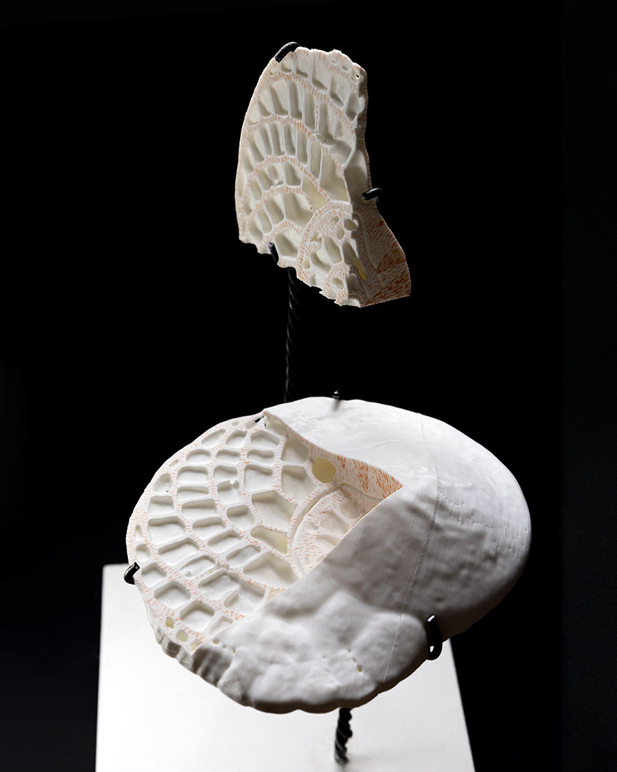
Heterostegina sp., 2023
3D print in PLA
34 × 22 × 18 cm
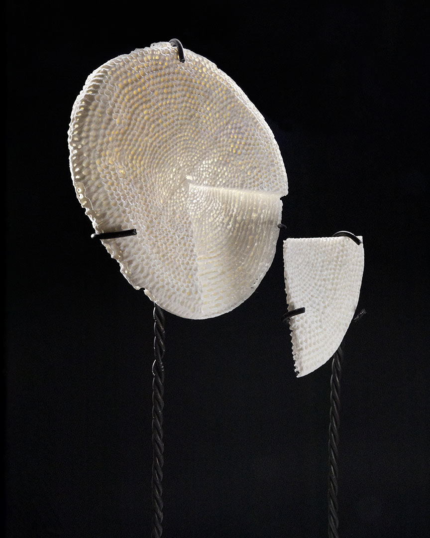
Marginopora sp., 2023
3D print in PLA
37 × 17 × 21.5 cm
To address this, the artists collaborated with a team of researchers to create educational models that reveal the diverse microscopic morphology of two extant genera: Heterostegina and Marginopora.
Their work combines multiple approaches and techniques, beginning with scientific dissemination through realistic educational sculptures. Using computed tomography (conducted by Adrián Belarra and Dr. Margarita Chevalier, UCM) and 3D rendering, they produced printed models of foraminifera based on specimens from Dr. Herrero’s collection (UCM). These forms were then reinterpreted artistically through cyanotypes. The sculptures and cyanotypes complement one another: the cuts made in the former help viewers understand the cross-sections depicted in the latter.
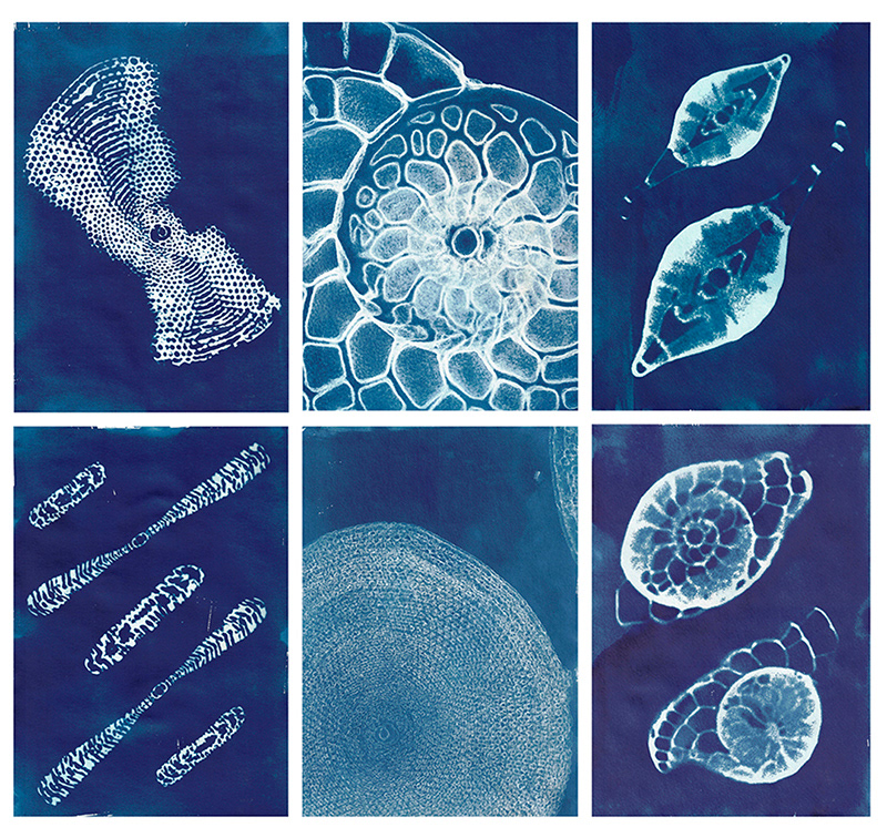
Gigaforam and Megaforam. Oblique axial and equatorial sections, 2023
Center column: Gigaforam Rx and Megaforam Rx, 2023
Based on X-ray effect renderings
Cyanotype on paper. 29.5 × 20.8 cm (each)
Foraminifera host microalgae that perform photosynthesis, aiding both shell formation and energy production. This inspired the use of cyanotype—a chemical process that harnesses sunlight to create Prussian blue images—referencing the first illustrated book printed entirely with photographs, Photographs of British Algae: Cyanotype Impressions (Atkins, 1843). In that pioneering work, the author combined scientific research, technological experimentation, and artistic expression to represent botanical specimens.